Blank Diagram Of A Long Bone - Unit 3 Part 1 Long Bone Diagram / Helps keep bones light in weight epiphyseal line line showing where growth plate used to be.
Blank Diagram Of A Long Bone - Unit 3 Part 1 Long Bone Diagram / Helps keep bones light in weight epiphyseal line line showing where growth plate used to be.. They support the body structurally, protect our vital organs, and allow us to move. This diagram makes it easier for one to display many potential causes for a specific effect or problem. There are four main categories of bones: The other primary skeletal component of. Sectional diagram of a long bone.
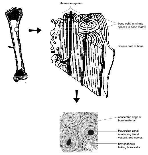
To view a high res version of an image click on the thumbnails. The hard cortical tissue can be invaded by cells that destroy the bone, called osteoclasts, only to have new bone laid down by secondary osteoblasts. Contains blood vessels, nerves, & lymph vessels. Skeletal diagrams are tools used by students to learn and study all 206 bones this number can vary from person to person of the human body. There is a printable worksheet available for download here so you can take the quiz with pen and paper.

Thick, fibrous membrane that covers the outside of a bone;
If it isn't present in your bone, draw a diagram in the blank box below to show the usual location of it. This diagram determines the possible causes of a specific event or problem. (c) identify one lamella on diagram a by using a bracket and label (the concentric ellae would be difficult to color without confusing other structures) Your diagram must take up at least half a page. Female pelvic bone anatomy images. Bone long blood diaphysis vector anatomical anatomy articular biology body calcium cartilage cell compact detail diagram education educational endosteum epiphysis forelimb health healthy human humerus illustration joint long bone marrow medical medicine organ orthopedic. Long bones — a subtype of bones — are longer than they are wide. Human anatomy for muscle, reproductive, and skeleton. Rebuilds the body's capacity to produce healthy cells. They are one of five types of bones: Skeletal diagrams are tools used by students to learn and study all 206 bones this number can vary from person to person of the human body. A long bone has two parts: The mineral calcium phosphate hardens this framework, giving it strength.
To view a high res version of an image click on the thumbnails. Enter the appropriate letter in the space provided. Related posts of diagram of of a long bone. We cover the diaphysis, the epiphysis, spongy and. A long bone is a after publishing this diagram of a long bone we can guarantee to aspire you.

Enter the appropriate letter in the space provided.
The long bones are those that are longer than they are wide. Long, short, irregular, and flat. Just print off and cut out. To view a high res version of an image click on the thumbnails. Related posts of diagram of of a long bone. The structure of a long bone allows for the best visualization of all of the parts of a bone (figure 1). Long bones, especially the femur and tibia, are subjected to most of the load during daily activities and they are crucial for skeletal mobility. Helps keep bones light in weight epiphyseal line line showing where growth plate used to be. This diagram makes it easier for one to display many potential causes for a specific effect or problem. Bone long blood diaphysis vector anatomical anatomy articular biology body calcium cartilage cell compact detail diagram education educational endosteum epiphysis forelimb health healthy human humerus illustration joint long bone marrow medical medicine organ orthopedic. A = epiphysis b = diaphysis c = articular cartilage d = periosteum f = compact bone g = medullary cavity (yellow marrow) h = endosteum j = epiphyseal line (growth plate). Clavicle diagram labeled anatomy human bones skeleton shoulder body wiring bone studyblue physiology flashcards system skeletal anatomia medical axial upper. This diagram determines the possible causes of a specific event or problem.
A = epiphysis b = diaphysis c = articular cartilage d = periosteum f = compact bone g = medullary cavity (yellow marrow) h = endosteum j = epiphyseal line (growth plate). Sectional diagram of a long bone. Long bones, especially the femur and tibia, are subjected to most of the load during daily activities and they are crucial for skeletal mobility. There is a printable worksheet available for download here so you can take the quiz with pen and paper. Bone marrow is the soft, highly vascular and flexible connective tissue within bone cavities.
af_3939 labeled diagram of the clavicle wiring diagram.
The long bones are those that are longer than they are wide. Also, they provide an environment for bones are mostly made of the protein collagen, which forms a soft framework. Bone marrow is the soft, highly vascular and flexible connective tissue within bone cavities. Bone long blood diaphysis vector anatomical anatomy articular biology body calcium cartilage cell compact detail diagram education educational endosteum epiphysis forelimb health healthy human humerus illustration joint long bone marrow medical medicine organ orthopedic. A = epiphysis b = diaphysis c = articular cartilage d = periosteum f = compact bone g = medullary cavity (yellow marrow) h = endosteum j = epiphyseal line (growth plate). In this video we discuss the parts of a long bone and some of the functions of each of those bone parts. Your diagram must take up at least half a page. This diagram makes it easier for one to display many potential causes for a specific effect or problem. Long, short, irregular, and flat. They support the body structurally, protect our vital organs, and allow us to move. Pobierz tę ilustrację wektorową diagram of a. Human anatomy for muscle reproductive and skeleton. Bone structure | anatomy and physiology i a typical long bone shows the gross anatomical characteristics of bone.


Komentar
Posting Komentar